When it comes to battling skin cancer, precision is key. Mohs surgery, a highly specialized procedure named after its inventor Dr. Frederic E. Mohs, stands out as one of the most effective treatments for certain types of skin cancer, particularly those with high recurrence rates or located in cosmetically sensitive areas. What makes Mohs surgery unique is its ability to meticulously remove cancerous tissue layer by layer, sparing healthy surrounding tissue. But how do dermatologists ensure they’ve eradicated all cancer roots during this intricate process?
Understanding the Process
Before delving into how dermatologists detect cancer roots during Mohs surgery, let’s first grasp the basics of this procedure. Mohs surgery involves the sequential removal of thin layers of cancerous skin tissue and immediate microscopic examination of each layer until no cancer cells remain. “Basically, we want to ensure that nothing is visible under the microscope and clear margins are obtained around the tumor that is removed,” explains Dr. Adam Mamelak, board-certified Dermatologist and Micrographic Dermatologic Surgeon. This precise approach maximizes the preservation of healthy tissue while minimizing the risk of cancer recurrence.
Spotting the Culprit: Cancer Roots
Cancer roots, also known as tumor “roots” or “fingers,” are invasive projections of cancer cells that extend beyond the visible tumor. These roots can infiltrate surrounding tissues and may not be readily apparent to the naked eye. However, dermatologists trained in Mohs surgery possess the expertise and tools necessary to identify these microscopic extensions during the procedure.
Microscopic Examination: The Key to Detection
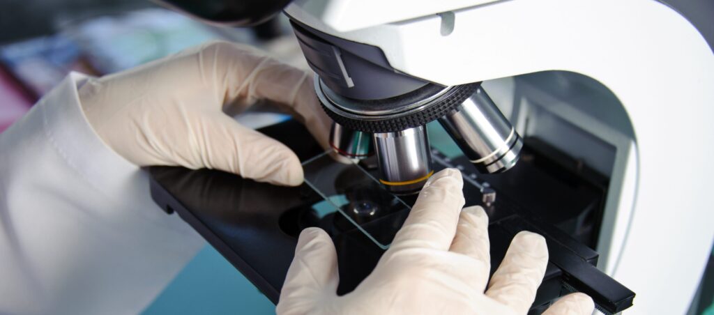
The cornerstone of Mohs surgery lies in its reliance on microscopic examination of tissue samples. After each layer of tissue is removed, it is meticulously examined under a microscope by the dermatologist. This process allows for real-time assessment of the margins of the excised tissue, enabling the detection of any remaining cancer cells or roots.
Staining Techniques Enhancing Visibility
To enhance the visibility of cancer cells and roots under the microscope, dermatopathologists and Mohs surgeons may employ various staining techniques. “Hematoxylin and eosin, and Toluidine blue are the most common stains used in Mohs surgery,” explains Dr. Mamelak. “Although immunohistochemistry, which involves the use of antibodies to highlight specific cellular components or proteins associated with cancer cells, is another approach we use in certain situations.” These stains can help differentiate between cancerous and healthy tissue, aiding in the identification of residual cancer roots.
Expertise and Experience Matter
Beyond the technical aspects of Mohs surgery, the expertise and experience of the dermatologist play a pivotal role in identifying cancer roots. Dermatologists trained in Mohs surgery undergo rigorous training and certification, equipping them with the knowledge and skills needed to navigate the complexities of skin cancer removal. Their keen understanding of tumor behavior and histopathology allows them to interpret microscopic findings accurately and make informed decisions during surgery.
The Importance of Complete Excision
Detecting and removing all cancer roots is crucial for achieving optimal outcomes in Mohs surgery. By meticulously examining tissue layers and ensuring complete excision of cancerous cells, dermatologists can significantly reduce the risk of cancer recurrence and minimize the need for additional treatments or surgeries.
Empowering Patients Through Education
As advocates for skin health, dermatologists play a vital role in educating patients about skin cancer prevention, detection, and treatment options. By raising awareness about Mohs surgery and its efficacy in treating skin cancer, dermatologists empower patients to make informed decisions about their healthcare and take proactive steps to protect their skin.
In conclusion, Mohs surgery stands as a gold standard in skin cancer treatment, offering unparalleled precision and effectiveness. Through meticulous microscopic examination and expertise in identifying cancer roots, dermatologists perform a delicate dance between preserving healthy tissue and eradicating cancerous cells. By shining a light on the invisible world of cancer roots, dermatologists continue to make significant strides in the fight against skin cancer, ensuring brighter and healthier futures for their patients.

